
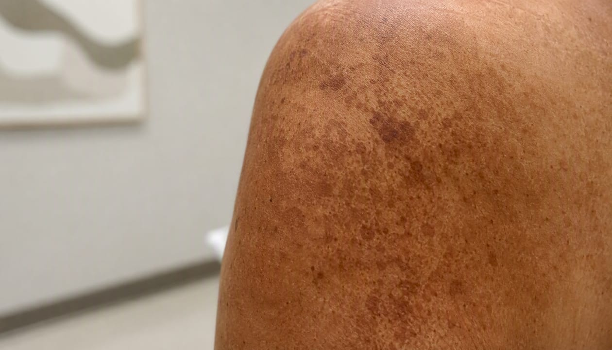
Why Sun Damage Causes Dark Spots: A Clear Explanation
Elizabeth Vogstrom, PA-C
evoyouthful Clinical Team
Discover why sun damage leads to dark spots and learn effective prevention strategies to protect your skin from UV and visible light.
Why Sun Damage Causes Dark Spots: A Clear Explanation
Sun exposure causes dark spots because UV and visible light stimulate melanocytes, the pigment-producing cells in your skin, to overproduce melanin in localized areas. Three mechanisms drive this process:
- Melanocyte activation: UV radiation signals melanocytes to ramp up melanin synthesis as a protective response, but that response is often uneven, creating concentrated patches of color.
- Inflammation-driven pigment: UV damage triggers an inflammatory cascade that further stimulates pigment production, a process known as post-inflammatory hyperpigmentation (PIH).
- Cumulative structural change: Repeated exposure degrades collagen and elastin (solar elastosis), altering the skin’s architecture in ways that make discoloration more persistent and harder to treat.
Two practical implications follow from this. First, prevention requires more than a standard sunscreen. Broad-spectrum protection against both UV and visible light is the most effective long-term strategy. Second, any spot that changes in size, shape, or color warrants a professional evaluation to rule out something more serious than a cosmetic concern. According to the Mayo Clinic, sunspots are common and benign, but changing lesions should always be assessed by a clinician.
Key Takeaways
UV and visible light cause dark spots by triggering melanocyte overactivation, inflammation, and cumulative structural skin damage that makes discoloration persistent without consistent photoprotection and targeted treatment.
| Point | Details |
|---|---|
| Core mechanism | UV and visible light stimulate melanocytes, trigger inflammation, and cause structural skin changes that produce focal hyperpigmentation. |
| Prevention is foundational | Broad-spectrum sunscreen with iron oxide, applied daily, is the most effective strategy for stopping new spots and preserving treatment results. |
| Spot type determines treatment | Solar lentigines, PIH, melasma, and freckles respond differently; accurate diagnosis before any procedure prevents worsening pigmentation. |
| Structural damage is not fully reversible | Treatments improve appearance significantly, but solar elastosis and collagen loss from chronic UV exposure cannot be fully restored. |
| evoyouthful’s approach | A full skin assessment and 3D imaging before any treatment ensures the right procedure for your skin type and minimizes PIH risk. |
Table of Contents
- Why sun damage causes dark spots: the biology behind it
- What types of dark spots does sun exposure actually cause?
- Who is most likely to develop sun-related dark spots?
- Do sunspots go away on their own?
- How to prevent new sun-related dark spots from forming
- Safe clinical and topical treatments to fade dark spots
- When should you see a dermatologist about a dark spot?
- What most patients get wrong about sun spots
- What we see in clinic and how patients typically progress
- Professional evaluation and personalized treatment at evoyouthful
- Sources
Why sun damage causes dark spots: the biology behind it
The connection between sun exposure and dark spots starts at the cellular level; understanding it helps you make smarter decisions about protection and treatment.
Which wavelengths do what. UVB rays (280–315 nm) are primarily responsible for sunburn and direct DNA damage. UVA rays (315–400 nm) penetrate more deeply, driving oxidative stress and long-term structural degradation. High-energy visible light, particularly wavelengths near the UVA boundary, can also produce DNA photoproducts and worsen pigmentation. This is why standard UV-only sunscreens sometimes fail to prevent dark spots entirely, as research published in the American Journal of Clinical Dermatology confirms.
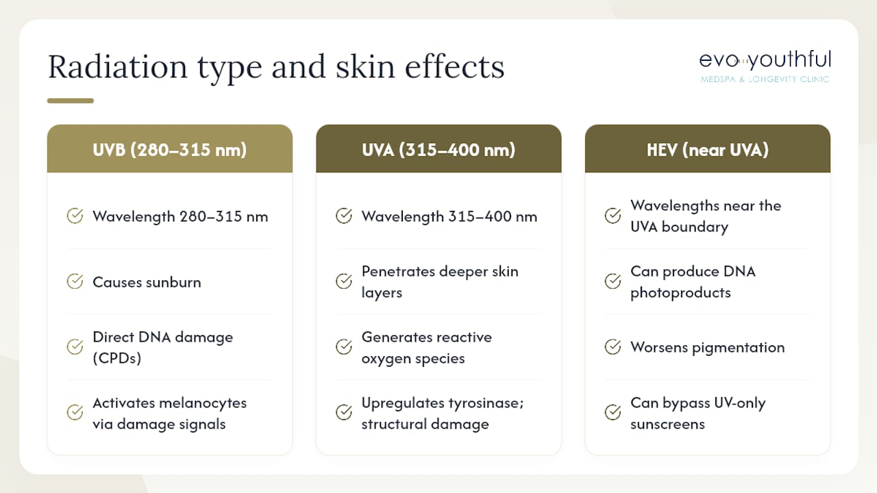
DNA damage and the pigmentation signal. When UVB strikes skin cells, it can cause cyclobutane pyrimidine dimers (CPDs), which are abnormal bonds between adjacent DNA bases. The cell’s repair machinery recognizes these lesions and, as part of its damage response, releases signals that activate melanocytes. This is a protective reflex, but it is imprecise. Melanocytes in certain areas respond more aggressively than others, producing focal overproduction of melanin rather than an even tan.
Oxidative stress and free radicals. UVA and infrared radiation generate reactive oxygen species (free radicals) that damage cell membranes, proteins, and DNA indirectly. This oxidative stress upregulates tyrosinase, the key enzyme in melanin synthesis, amplifying pigment production beyond what direct UV damage alone would cause. The result is a second, parallel pathway to hyperpigmentation that persists even on cloudy days or behind glass.
Inflammation as a pigment amplifier. UV exposure triggers the release of prostaglandins, cytokines, and other inflammatory mediators. These molecules stimulate melanocyte hyperactivity and, after the acute inflammation resolves, can leave behind excess pigment in the dermis and epidermis. The Merck Manual notes that UV radiation damages skin through direct DNA injury, oxidative damage, and inflammation together, and that these mechanisms collectively promote pigmentation changes and increase the risk of genetic mutations in skin cells.
Melanosome transfer and why spots persist. Melanocytes produce melanin inside organelles called melanosomes and transfer them to surrounding keratinocytes (the cells that form the outer skin layers). When melanocyte hyperfunction continues over years, the transfer becomes dysregulated. Melanosomes accumulate unevenly, and in areas of chronic exposure, the skin’s structural changes from solar elastosis make it harder for normal cell turnover to clear the excess pigment. That is why a sunspot from a decade of outdoor work does not simply fade the way a fresh bruise does.
What types of dark spots does sun exposure actually cause?
Not every brown spot on your skin has the same origin, and the distinction matters for choosing the right treatment.
Solar lentigines (sunspots) are flat, well-defined brown macules that appear on chronically sun-exposed areas: the face, hands, forearms, and chest. They develop from long-term cumulative UV exposure rather than a single sunburn and tend to appear after age 40, though they can occur earlier in people with fair skin or high lifetime sun exposure. According to Healthline, solar lentigines are benign and are often treated for cosmetic reasons using topical agents and dermatologic procedures.
Ephelides (freckles) appear in childhood and adolescence, are strongly linked to genetics, and tend to fade in winter when UV exposure drops. They represent a normal variation in melanocyte response rather than a sign of damage, though they do indicate UV sensitivity.
Melasma is a hormonally influenced form of hyperpigmentation that UV exposure reliably worsens. It appears as larger, irregular patches, typically on the cheeks, forehead, and upper lip. Pregnancy, oral contraceptives, and hormonal fluctuations are common triggers. Melasma is notoriously difficult to treat because it tends to recur with any UV exposure.
Post-inflammatory hyperpigmentation (PIH) follows skin injury or inflammation, including acne, eczema, cuts, or even aggressive cosmetic procedures. UV exposure after the initial injury deepens the pigment. The Merck Manual notes that focal hyperpigmentation is most often postinflammatory and that people with darker skin tones are more susceptible to PIH, making careful treatment selection especially important.
Why identification matters before treatment. A solar lentigo and an early melanoma can look similar to the untrained eye. PIH responds differently to lasers than a solar lentigo does, and melasma can actually worsen with certain laser settings. Getting the diagnosis right first is not a formality; it directly determines whether a treatment helps or harms.
Who is most likely to develop sun-related dark spots?
Several factors shape your personal risk, and some of them are modifiable.
Cumulative UV dose is the single largest driver. People who have spent decades working outdoors, living at high altitude, or in high-reflectance environments (snow, water, sand) carry a heavier burden of sun-related skin changes. Tanning bed use adds significant UVA exposure on top of natural sunlight, accelerating the process.
Fitzpatrick skin type influences how risk presents. Lighter skin types (I–III) tend to develop solar lentigines and photoaging changes earlier. Darker skin types (IV–VI) have more natural UV protection from higher baseline melanin, but when hyperpigmentation does develop, it tends to be more pronounced and harder to treat because of the elevated PIH risk.
Hormonal factors play a meaningful role. Estrogen and progesterone sensitize melanocytes to UV stimulation, which is why melasma is far more common in women during pregnancy or while using oral contraceptives.

Medications and photosensitizers are an underappreciated risk factor. Certain antibiotics (doxycycline, tetracycline), diuretics, NSAIDs, and some herbal supplements increase UV sensitivity and can cause phototoxic reactions that leave focal hyperpigmentation behind. Phytophotodermatitis, caused by contact with plants like limes or parsley followed by sun exposure, is a classic example. The Merck Manual advises clinicians to screen for photosensitizing exposures when evaluating new spots.
Genetics determine baseline melanocyte reactivity, skin repair efficiency, and susceptibility to UV-induced DNA damage. A family history of melasma or early sunspots is a reliable predictor of personal risk.
Age compounds all of the above. As skin ages, melanocyte distribution becomes less uniform, cell turnover slows, and the skin’s ability to repair UV-induced DNA damage declines, making new spots more likely and existing ones more persistent.
Do sunspots go away on their own?
The honest answer is: it depends on the type of spot and how well you protect your skin going forward.
- PIH from acne or minor injury can fade over several months to a year with consistent sun protection and no further inflammation. The deeper the pigment sits in the dermis, the slower the resolution.
- Freckles often lighten noticeably in winter and with age as UV exposure decreases, though they rarely disappear entirely in people with a genetic predisposition.
- Solar lentigines are generally long-lasting without intervention. They may lighten slightly with strict sun avoidance, but most do not resolve on their own once established.
- Melasma is the most persistent. Without strict photoprotection and hormonal management, it recurs reliably after any treatment.
Structural sun damage, specifically the collagen and elastin breakdown of solar elastosis, is not fully reversible. Treatments can significantly improve the appearance of sun-damaged skin, but they do not restore the original tissue architecture. Research published in the American Journal of Clinical Dermatology confirms that chronic UV exposure causes photoaging and structural degradation that treatments can improve but not fully reverse.
The clearest takeaway: prevention is the most effective long-term strategy. Starting or strengthening a photoprotection routine now limits new spot formation and preserves the results of any treatment you pursue.
How to prevent new sun-related dark spots from forming
Start with broad-spectrum sunscreen, every day. A broad-spectrum SPF 30 or higher applied every morning, regardless of cloud cover, blocks both UVA and UVB. Apply an adequate amount to the face and exposed body areas. Reapply regularly when outdoors or after swimming or sweating.
Add a tinted sunscreen with iron oxide. Standard sunscreens do not block visible light, which contributes meaningfully to hyperpigmentation. Tinted formulas containing iron oxide provide that additional protection. The American Academy of Dermatology specifically recommends iron oxide-containing sunscreens for people prone to dark spots, and they have the added benefit of avoiding the white cast that mineral sunscreens can leave on darker skin tones.
Layer a topical antioxidant underneath. Vitamin C serum (L-ascorbic acid, 10–20%) applied before sunscreen neutralizes free radicals that UV and visible light generate, reducing the oxidative stress pathway to pigmentation. This combination is more protective than sunscreen alone.
Use physical barriers. UPF 50+ clothing, wide-brim hats, and UV-blocking sunglasses reduce cumulative dose significantly, especially for outdoor workers or those who spend extended time near reflective surfaces like water or snow.
Avoid peak sun hours and tanning beds. UV intensity is highest between 10 AM and 4 PM. Tanning beds deliver concentrated UVA exposure with no protective benefit, and their use is directly associated with accelerated photoaging and increased hyperpigmentation risk.
Pro Tip: If you take doxycycline, a diuretic, or certain herbal supplements, ask your prescriber about photosensitivity risk. Pairing a photosensitizing medication with unprotected sun exposure can cause hyperpigmentation that takes months to resolve.
Safe clinical and topical treatments to fade dark spots
Choosing the right treatment depends on the type of spot, your skin tone, and whether you want a topical approach, a clinical procedure, or a combination of both.
| Treatment | How it works | Typical time to results | Key risks |
|---|---|---|---|
| Hydroquinone (Rx) | Inhibits tyrosinase, reducing melanin synthesis | 8 weeks | Ochronosis with prolonged use; requires cycling |
| Retinoids (tretinoin) | Accelerates cell turnover, disperses melanin | 12 weeks | Irritation, initial photosensitivity |
| Azelaic acid | Selectively inhibits hyperactive melanocytes | 12 weeks | Mild stinging; generally well tolerated |
| Vitamin C (topical) | Antioxidant; inhibits tyrosinase | 8 weeks | Oxidizes quickly; formulation matters |
| Licorice extract | Glabridin inhibits tyrosinase | 8 weeks | Mild; suitable for sensitive skin |
| Chemical peels | Exfoliates pigmented layers | 4 sessions | PIH risk, especially in darker skin |
| Cryotherapy | Freezes and destroys pigmented cells | 1–3 sessions | Hypopigmentation risk |
| IPL / non-ablative laser | Targets melanin with selective photothermolysis | 3 sessions | PIH risk in skin of color; requires test spot |
| RF microneedling | Stimulates collagen remodeling; disperses pigment | 3–4 sessions | Moderate PIH risk; lower than ablative laser |
| Microneedling + topicals | Channels topical agents deeper for enhanced effect | 3 sessions | PIH risk if performed without proper assessment |
Combining therapies tends to work faster. DermNet NZ notes that combining topical prescription agents with resurfacing procedures generally achieves faster and more complete clearing than single modalities, though aggressive treatments carry a nontrivial risk of new dyschromia if applied without proper assessment. A prescription topical retinoid or hydroquinone used alongside a conservative resurfacing treatment is a common first-line combination.
Darker skin tones require extra caution. PIH risk is higher in Fitzpatrick types IV–VI, and any heat-based or ablative procedure should begin with a test spot at conservative settings. Collagen induction therapy via microneedling is often a safer starting point than laser for patients with higher PIH risk, particularly when combined with a topical brightening regimen.
Pro Tip: Never start an aggressive resurfacing procedure without a confirmed diagnosis. A spot that looks like a solar lentigo but has irregular borders or color variation needs a dermatologist’s assessment before any cosmetic treatment.
When should you see a dermatologist about a dark spot?
Most dark spots are benign, but certain features signal that a spot needs prompt medical evaluation rather than cosmetic care.
Red flags that warrant urgent assessment:
- A spot that has changed in size, shape, or color over weeks to months
- Irregular, asymmetric, or poorly defined borders
- Multiple colors within a single lesion (brown, black, red, or white areas)
- Bleeding, crusting, or ulceration without injury
- A rapidly growing lesion
- Any spot that itches, hurts, or feels different from surrounding skin
What to expect at the appointment. A dermatologist will perform a clinical exam and typically use dermoscopy, a magnified, polarized-light examination, to assess the lesion’s internal structure. Photo documentation creates a baseline for monitoring change over time. When malignancy cannot be excluded clinically, a biopsy is the standard next step.
What to bring. A timeline of when the spot appeared and how it has changed, photos if you have them, a list of all current medications and supplements (including any photosensitizers), and a record of prior treatments or products applied to the area.
Before any cosmetic procedure. A pre-procedure assessment should include a discussion of your skin type, PIH risk, and a test spot for any heat-based or ablative treatment. Informed consent for aesthetic procedures should include a clear explanation of the risk of post-procedural dyschromia, particularly for patients with darker skin tones or a history of PIH.
What most patients get wrong about sun spots
A common misconception is that only UVB rays matter for pigmentation because UVB causes sunburn. In practice, UVA penetrates more deeply, drives oxidative stress year-round, and is the primary driver of long-term photoaging and solar lentigines. UVA passes through window glass and remains intense even on overcast days, which is why daily sunscreen use matters even when you are not planning to be outdoors.
A second misconception: “I’ll treat the spots first and worry about sunscreen later.” Without strict photoprotection, any topical or procedural treatment is working against continued UV stimulation. The American Academy of Dermatology is direct on this point: preventing new pigmentation with photoprotection is the foundation of any dark spot management plan, and without it, active treatments are often undone by continued exposure.
A third: “all lasers are equally safe for my skin tone.” They are not. IPL and ablative lasers carry meaningfully higher PIH risk in darker skin tones than non-ablative or RF-based options. The right device, settings, and operator experience all determine whether a procedure clears a spot or creates a new one. This is precisely why a thorough pre-treatment assessment at a qualified clinic matters so much.
What we see in clinic and how patients typically progress
The patients who respond best to treatment share one thing: they commit to photoprotection from day one and maintain it throughout. The most common setback is inconsistent sunscreen use, which allows UV stimulation to continue working against whatever topical or procedural treatment is underway.
Two scenarios come up regularly. The first is a patient in their 30s or 40s dealing with PIH after acne or a prior procedure. With a consistent topical regimen (azelaic acid or a low-dose retinoid, paired with a tinted iron oxide sunscreen), most see meaningful improvement within three to four months. The second is a patient with long-standing solar lentigines on the hands or face who expects a single laser session to clear everything. In reality, a combination approach, starting with a topical prescription phase, followed by a conservative resurfacing procedure, then a maintenance regimen, produces the most durable results.
The typical clinic pathway at evoyouthful looks like this: a full skin assessment with photo documentation, a topical trial phase to stabilize pigment and reduce inflammation, a procedural option when appropriate (RF microneedling, Exion Clear, or a pigment-safe laser), and a long-term maintenance plan built around daily photoprotection and physician-curated skincare. Patients who follow this pathway and protect their results with sunscreen tend to hold their improvement well. Those who skip the maintenance phase often see gradual recurrence within a year.
For patients with uneven skin tone and texture concerns that go beyond pigmentation, the same assessment-first approach applies. Photoaging affects collagen and elastin as well as color, and addressing both together produces more complete results than targeting pigment alone.
Professional evaluation and personalized treatment at evoyouthful
Persistent or suspicious dark spots deserve more than an over-the-counter cream and a wait-and-see approach. At evoyouthful in Wayzata, MN, the starting point is always a thorough skin assessment, including advanced 3D imaging, to accurately identify the type of pigmentation, assess your skin type, and rule out anything that needs a dermatologist’s attention before any cosmetic treatment begins.

From there, the clinic offers a range of treatments matched to your specific skin type and goals: Exion Clear RF microablation for resurfacing and pigment reduction, RF microneedling, PRF injections for skin renewal, physician-curated topical regimens, and pigment-safe laser and IPL options selected with your Fitzpatrick type in mind. For patients whose hyperpigmentation has a hormonal component, the team can also discuss how hormonal imbalance may be contributing to melasma or persistent pigmentation changes.
Your initial consultation includes a full skin assessment, photo documentation, a personalized treatment plan with a realistic timeline, and a clear discussion of what each option involves. To schedule your consultation, visit Evoyouthful or call the Wayzata clinic directly.
Sources
- Photoaging: Current Concepts on Molecular Mechanisms, Prevention, and Treatment | American Journal of Clinical Dermatology | Springer Nature Link
- How to fade dark spots in darker skin tones
- Overview of Effects of Sunlight - Dermatology - Merck Manual Professional Edition
- Sun damage - Mayo Clinic
- Removing Sunspots on Face: Treatment and At-Home Remedies
- Solar lentigo — DermNet NZ
This article is general information, not a substitute for advice from a qualified doctor. Consult a qualified healthcare professional about your own circumstances before acting on anything here.
Recommended
- Acne Scarring | Treatments in Wayzata, MN | evoyouthful Wayzata, MN
- Uneven Skin Tone & Texture | Treatments in Wayzata, MN | evoyouthful Wayzata, MN
- Fine Lines & Wrinkles | Treatments in Wayzata, MN | evoyouthful Wayzata, MN
- Skin Hydration & Plumpness | Treatments in Wayzata, MN | evoyouthful Wayzata, MN
Ready to Start?
Schedule a Consultation
Have questions about what you just read? Our team is here to help.